
Question 1
Which statement about centrioles is correct?
A. Centrioles are formed from microtubules
B. Centrioles are only found in plant cells
C. Centrioles are found in the nucleus
D. Centrioles are involved in the synthesis of ATP
Easy
Mark as Complete
Mark Scheme
Question 2
Which statement is correct about bacteria?
A. Bacterial cells contain circular DNA
B. Bacterial cells contain a nuclear envelope
C. Bacterial cells contain `80`S ribosomes
D. Bacterial cells contain mitochondria
Easy
Mark as Complete
Mark Scheme
Question 3
Identify which option correctly states the two structures that all viruses consist of.
A. DNA/RNA and a cell surface membrane
B. DNA/RNA and a cell wall
C. a protein capsid and a cell surface membrane
D. a protein capsid and DNA/RNA
Easy
Mark as Complete
Mark Scheme
Question 4
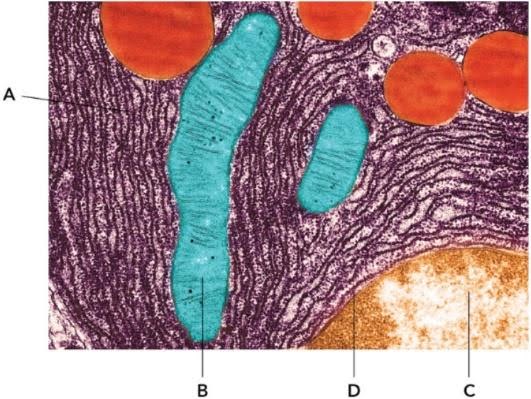
The figure below shows organelles on a transmission electron micrograph of part of a cell from the pancreas. Use the list provided to identify parts A–D.
Easy
Mark as Complete
Mark Scheme
Question 5
Which of the following shows the organelles in order of size, starting with the smallest?
A. lysosome → mitochondria → nucleus → ribosome
B. ribosome → lysosome → mitochondria → nucleus
C. nucleus → ribosome → lysosome → mitochondria
D. mitochondria → nucleus → ribosome → lysosome
Medium
Mark as Complete
Mark Scheme
Question 6
In vertebrates, beating cilia are also found on the epithelial cells of the oviduct (the tube connecting the ovary to the uterus). Suggest what function cilia have in the oviduct.
Cilia in the oviduct beat in order to move the egg from the ovary to the uterus. (It may be fertilised along the way.)
Medium
Mark as Complete
Mark Scheme
Question 7
List the structural features that prokaryotic and eukaryotic cells have in common. Briefly explain why each of the structures you have listed is essential.
Medium
Mark as Complete
Mark Scheme
Question 8
The figure is a diagram based on an electron micrograph of a secretory cell from the pancreas. This type of cell is specialised for secreting (exporting) proteins. Some of the proteins are digestive enzymes of the pancreatic juice. The cell is very active, requiring a lot of energy. The arrows A, B, C and D show the route taken by the protein molecules. Note that arrow A is shown magnified in a separate diagram.
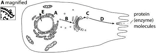
a. Describe briefly what is happening at each of the stages A, B, C and D.
b. Arrow E shows the path of a molecule or structure leaving the nucleus through the nuclear envelope. Name one molecule or structure which leaves the nucleus by route E.
c. Name the molecule which leaves the mitochondrion in order to provide energy for the cell.
Medium
Mark as Complete
Mark Scheme
Question 9
All cells are surrounded by a cell surface membrane, and also contain many other membranes within them. Researchers estimated the total quantity of membranes in `20` liver cells and `20`exocrine pancreas cells, and then calculated the percentage of these membranes in all the different membrane-containing structures in the cells. Their results are shown in the table.
| Source of membrane | Mean percentage of all membranes | |
| Liver cells | Exocrine pancreas cells | |
| cell surface membrane | 1.8 | 4.7 |
| mitochondrial membrane | 39.4 | 22.3 |
| nuclear membrane | 0.5 | 0.7 |
| rough endoplasmic reticulum | 33.4 | 61.9 |
| smooth endoplasmic reticulum | 16.3 | 0.1 |
| Golgi apparatus | 7.9 | 10.3 |
| lysosomes | 0.4 | 0 |
| other small vesicles | 0.3 | 0 |
a. Explain why we cannot use these results to draw the conclusion that the mean quantity of cell surface membrane in liver cells is less than that in exocrine pancreas cells.
b. Which of the sources of membranes listed in the table are made up of two membranes (an envelope)?
c. Using the data in the table, state the organelle that contains the greatest mean percentage of membrane in:
i. liver cells
ii. pancreas cells
d. Liver cells have a wide variety of functions in metabolism, including synthesising proteins, breaking down toxins, synthesising cholesterol and producing bile. Exocrine pancreas cells have a single main role, which is the production and secretion of digestive enzymes. Use this information to suggest explanations for the differences between the percentages for mitochondria and rough endoplasmic reticulum in the liver cells and the pancreas cells.
Hard
Mark as Complete
Mark Scheme
Question 10
The photograph shows a micrograph of parts of two cells from the small intestine of a mammal. The structures along the facing surfaces of the two cells are microvilli.
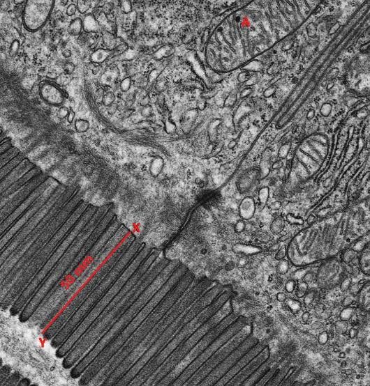
a. State the type of microscope that was used to obtain this micrograph. Give a reason for your answer.
b. Identify organelle A.
c.
i. The magnification of the micrograph is `× 12 500`. The length XY in micrograph is `50` mm. Calculate the actual length of the microvillus between points X and Y. Show your working.
ii. Microvilli greatly increase the surface areas of the cells. Suggest why the cells lining the small intestine have microvilli.
Hard
Mark as Complete
Mark Scheme
Question 1
Which statement about centrioles is correct?
A. Centrioles are formed from microtubules
B. Centrioles are only found in plant cells
C. Centrioles are found in the nucleus
D. Centrioles are involved in the synthesis of ATP
Answer: A
A. Correct: Centrioles are made of microtubules. Specifically, a centriole is a hollow cylinder made from a ring of short microtubules arranged in nine triplets of microtubules.
B. Incorrect: Centrioles are typically found in animal cells. They are absent from most plant cells, flowering plants and fungi, or that plant cells do not contain centrosomes (which contain centrioles).
C. Incorrect: Centrioles are located outside the nucleus. They are typically found as a pair within a region called the centrosome, and two centrioles often lie at right angles to each other.
D. Incorrect: The function of centrioles is primarily related to cell division, specifically spindle formation in animal cells, and the formation and movement of cilia and flagella. Mitochondria are the site of ATP synthesis. Chloroplasts also produce ATP.
Question 2
Which statement is correct about bacteria?
A. Bacterial cells contain circular DNA
B. Bacterial cells contain a nuclear envelope
C. Bacterial cells contain `80`S ribosomes
D. Bacterial cells contain mitochondria
Answer: A
A. Correct: Bacterial cells, which are prokaryotic cells, contain their main genetic material as a single circular DNA molecule that is free in the cytoplasm. This main DNA loop is typically not associated with proteins like histones. Many bacteria may also have smaller circular pieces of DNA called plasmids.
B. Incorrect: Bacterial cells are prokaryotic and lack a true nucleus. They do not have a nuclear membrane or envelope. The circular DNA is found in a region called the nucleoid, which also contains proteins and small amount of RNA. Prokaryotic cells generally lack membrane-bound organelles in their cytoplasm.
C. Incorrect: Bacterial cells contain smaller `70`S ribosomes. Eukaryotic cells, such as animal and plant cells, have larger `80`S ribosomes in their cytoplasm. (Note that the mitochondria and chloroplasts within eukaryotic cells do contain `70`S ribosomes, which is one piece of evidence supporting the endosymbiotic theory of their origin from bacteria.)
D. Incorrect: Bacterial cells, being prokaryotic, lack membrane-bound organelles. Mitochondria are membrane-bound organelles found in eukaryotic cells.
Question 3
Identify which option correctly states the two structures that all viruses consist of.
A. DNA/RNA and a cell surface membrane
B. DNA/RNA and a cell wall
C. a protein capsid and a cell surface membrane
D. a protein capsid and DNA/RNA
Answer: D
A. Incorrect: Viruses are disease-causing agents, rather than ‘organisms’. They do not have cellular structures, hence do not have a cell surface membrane.
B. Incorrect: Viruses do not have cellular structures and do not have a cell wall. Cell walls are found in prokaryotic cells (like bacteria) and some eukaryotic cells (like plants and fungi).
C. Incorrect: While viruses do have a protein coat called a capsid, they lack a cell surface membrane.
D. Correct: Viruses consist of a core of nucleic acid (either DNA or RNA) surrounded by a protein coat, called a capsid. Some viruses may have an additional outer layer, such as an envelope, but the core nucleic acid and the protein capsid are the fundamental components present in all viruses.
Question 4
The figure below shows organelles on a transmission electron micrograph of part of a cell from the pancreas. Use the list provided to identify parts A–D.
A. rough endoplasmic reticulum (RER). The network of membranes studded with small dark dots (ribosomes) indicates the RER. This structure is key in protein synthesis, especially abundant in secretory cells like those in the pancreas.
B. mitochondrion. The rod-shaped structure with internal cristae (folds) is mitochondrion (plural: mitochondria). It is the site of the aerobic stages of respiration and the site of the synthesis of much ATP.
C. nuclear envelope. The outer double membrane surrounding the nucleus is the nuclear envelope. It regulates the flow of materials between the nucleus and cytoplasm.
D. nucleus. The largest organelle just inside the nuclear envelope represents the nucleus itself. It contains the cell's genetic material (DNA) and controls gene expression.
Question 5
Which of the following shows the organelles in order of size, starting with the smallest?
A. lysosome → mitochondria → nucleus → ribosome
B. ribosome → lysosome → mitochondria → nucleus
C. nucleus → ribosome → lysosome → mitochondria
D. mitochondria → nucleus → ribosome → lysosome
Answer: B
Ribosomes are the smallest and most numerous of the cell organelles. Their diameter is approximately `25 nm`. (Note that bacterial ribosomes are `70`S ribosomes, slightly smaller than the `80`S ribosomes of eukaryotes.)
Lysosomes are "little membrane-bound packages", containing enzymes. They are significantly larger than ribosomes but still smaller than mitochondria and the nucleus.
Mitochondria are relatively large organelles, typically `0.1-1.5 μm` wide and `3.0-10.0 μm` long. Their diameter is about `1 μm`.
The nucleus is the largest organelle in eukaryotic cells. It is typically `10-20 μm` in diameter, or about `5-10 μm` in diameter.
Comparing these sizes, ribosomes (nm range) are significantly smaller than mitochondria and the nucleus (μm range). The nucleus is larger than mitochondria. Lysosomes are generally smaller than mitochondria and the nucleus.
So the correct order is ribosome → lysosome → mitochondria → nucleus.
Question 6
In vertebrates, beating cilia are also found on the epithelial cells of the oviduct (the tube connecting the ovary to the uterus). Suggest what function cilia have in the oviduct.
Cilia in the oviduct beat in order to move the egg from the ovary to the uterus. (It may be fertilised along the way.)
Cilia are whip-like, beating extensions of many eukaryotic cells. They can cause the movement of fluid across the cell surface or propel cells through fluid. The cilia in the oviduct beat in order to move the egg from the ovary towards the uterus. This action provides the propulsion needed to carry the egg cell along the tube. This movement is important for the process of reproduction. Fertilisation may occur along the way as the egg is moved down the oviduct.
Hormones like oestrogen can influence the cilia in the oviduct, causing the development of more, larger, and more active cilia. These changes help prepare the oviduct for the arrival of the oocyte, which will be moved along the oviduct by the cilia.
Question 7
List the structural features that prokaryotic and eukaryotic cells have in common. Briefly explain why each of the structures you have listed is essential.
Prokaryotic and eukaryotic cells share some fundamental structural features and essential functions, despite significant differences in complexity and other organelles.
The structural features common to both prokaryotic and eukaryotic cells are the cell surface membrane, cytoplasm, ribosomes, and DNA.
Cell surface membrane: This membrane surrounds all cells, including both eukaryotes and prokaryotes. It acts as a partially permeable barrier between the cell's internal environment and the external environment. Its essential function is to control the movement of substances into and out of the cell, regulating exchange. This control is necessary to maintain a specific internal environment different from the surroundings. Without it, the chemistry of life would mix with the environment, making a separate chemistry of life impossible.
Cytoplasm: This is the material within the cell surface membrane. It is the site of metabolic activity and contains biochemicals in solution. Many of the chemical reactions essential for life occur within the cytoplasm.
Ribosomes: These are very small organelles found in both prokaryotic and eukaryotic cells. In eukaryotic cells, ribosomes in the cytosol are `80`S, while those in mitochondria and chloroplasts are `70`S. Prokaryotic ribosomes are `70`S. Ribosomes are not surrounded by a membrane. Their essential function is to be the site of protein synthesis, an activity vital for all cells.
DNA: This is the genetic material found in all cells. In eukaryotic cells, DNA is typically linear and located within the nucleus, although mitochondria and chloroplasts also contain circular DNA. In prokaryotic cells, DNA is typically circular and located freely in the cytoplasm (in a region called the nucleoid). Its essential functions include containing the information or instructions that control the cell's activities, particularly controlling protein synthesis. DNA is also capable of replicating itself, which is necessary for cell division and the formation of new cells.
Note that the cell wall and flagellum are not structural features common to all prokaryotic and eukaryotic cells. Cell walls are present in plant, fungal, and prokaryotic cells, but are absent in animal cells, which are eukaryotic. Flagella are present in some prokaryotes and some eukaryotic cells (like gametes or certain single-celled organisms), but not all eukaryotes possess them.
Question 8
The figure is a diagram based on an electron micrograph of a secretory cell from the pancreas. This type of cell is specialised for secreting (exporting) proteins. Some of the proteins are digestive enzymes of the pancreatic juice. The cell is very active, requiring a lot of energy. The arrows A, B, C and D show the route taken by the protein molecules. Note that arrow A is shown magnified in a separate diagram.
a. Describe briefly what is happening at each of the stages A, B, C and D.
b. Arrow E shows the path of a molecule or structure leaving the nucleus through the nuclear envelope. Name one molecule or structure which leaves the nucleus by route E.
c. Name the molecule which leaves the mitochondrion in order to provide energy for the cell.
a. The figure depicts a pancreatic cell specialized for secreting (exporting) proteins, some of which are digestive enzymes found in pancreatic juice. This process requires significant energy, hence such cells are very active and contain many mitochondria.
Stage A: Protein is made on the ribosome and is moving into the rough ER. Ribosomes are the sites of protein synthesis. Ribosomes that are attached to the membranes of the endoplasmic reticulum (forming rough endoplasmic reticulum, RER) are the site of synthesis for proteins that are subsequently secreted. As proteins are made on these ribosomes, they enter the sacs or cisternae of the RER.
Stage B: The rough ER buds off small vesicles. These vesicles contain the proteins. The vesicles then fuse to form the Golgi apparatus or fuse with the Golgi apparatus. As a result, the protein moves into the Golgi apparatus. Inside the Golgi apparatus, the protein may be modified or processed, for example, by adding sugars to form glycoproteins. The Golgi apparatus collects and processes molecules from the RER.
Stage C: The Golgi apparatus buds off Golgi vesicles. These vesicles are also known as secretory vesicles because they contain materials to be secreted from the cell.
Stage D: The Golgi vesicles travel to the cell surface membrane. Here, the vesicle(s) fuses with the cell surface membrane. This fusion process releases the contents, the protein or enzyme, leaves the cell. This specific mechanism of bulk transport out of the cell is called exocytosis, also referred to as secretion. Exocytosis is an active process that requires ATP.
b. Regarding the arrow E, a molecule or structure that leave the nucleus through the nuclear envelope (in the context of protein secretion) is:
Messenger RNA (mRNA). mRNA is transcribed from DNA in the nucleus and carries the genetic code for protein synthesis to the cytoplasm.
or
Ribosomes. Ribosomes are manufactured in the nucleolus, which is located inside the nucleus. Ribosomes are composed of ribosomal RNA (rRNA) and protein. They function as the site of protein synthesis in the cytoplasm.
c. The molecule which leaves the mitochondrion in order to provide energy for the cell is adenosine triphosphate (ATP).
Mitochondria are the sites of aerobic respiration. Aerobic respiration releases energy, and the energy released from processes like the breakdown of glucose is used to make ATP. ATP synthesis occurs in mitochondria. ATP is produced in the mitochondrial matrix by ATP synthase can be used for all the energy-requiring reactions of the cell, both inside and outside the mitochondrion.
ATP is the universal energy currency molecule of cells. It acts as the immediate source of energy in a cell. ATP is a small, soluble molecule and can diffuse or move easily around the cell. ATP diffuses to the part of the cell that needs energy. When energy is required by the cell, ATP is broken down (hydrolysed) into ADP and inorganic phosphate, releasing energy. This released energy is then used for various cellular activities.
Question 9
All cells are surrounded by a cell surface membrane, and also contain many other membranes within them. Researchers estimated the total quantity of membranes in `20` liver cells and `20`exocrine pancreas cells, and then calculated the percentage of these membranes in all the different membrane-containing structures in the cells. Their results are shown in the table.
| Source of membrane | Mean percentage of all membranes | |
| Liver cells | Exocrine pancreas cells | |
| cell surface membrane | 1.8 | 4.7 |
| mitochondrial membrane | 39.4 | 22.3 |
| nuclear membrane | 0.5 | 0.7 |
| rough endoplasmic reticulum | 33.4 | 61.9 |
| smooth endoplasmic reticulum | 16.3 | 0.1 |
| Golgi apparatus | 7.9 | 10.3 |
| lysosomes | 0.4 | 0 |
| other small vesicles | 0.3 | 0 |
a. Explain why we cannot use these results to draw the conclusion that the mean quantity of cell surface membrane in liver cells is less than that in exocrine pancreas cells.
b. Which of the sources of membranes listed in the table are made up of two membranes (an envelope)?
c. Using the data in the table, state the organelle that contains the greatest mean percentage of membrane in:
i. liver cells
ii. pancreas cells
d. Liver cells have a wide variety of functions in metabolism, including synthesising proteins, breaking down toxins, synthesising cholesterol and producing bile. Exocrine pancreas cells have a single main role, which is the production and secretion of digestive enzymes. Use this information to suggest explanations for the differences between the percentages for mitochondria and rough endoplasmic reticulum in the liver cells and the pancreas cells.
a. The table shows the mean percentage of the total quantity of membrane contributed by each listed membrane-containing structure within either liver cells or exocrine pancreas cells. However, the data does not provide information about the actual total quantity of membrane in each cell type.
Hence, it is impossible to conclude that the mean quantity of cell surface membrane in liver cells is less than that in exocrine pancreas cells.
b. The sources of membranes that are made up of two membranes (an envelope) are the mitochondrial membranes and the nuclear membrane.
c. Based on the data provided in the table, the organelle that contains the greatest mean percentage of membrane in:
i. Liver cells is mitochondrial membrane `(39.4%)`.
ii. Pancreas cells is rough endoplasmic reticulum `(61.9%)`.
d. The table shows that in liver cells, mitochondrial membranes constitute the largest mean percentage of all membranes at `39.4%`, while rough endoplasmic reticulum (RER) accounts for `33.4%`. In contrast, exocrine pancreas cells have a much higher mean percentage of RER membranes `(61.9%)` and a lower percentage of mitochondrial membranes `(22.3%)`.
These differences in membrane percentages can be explained by the primary functions of each cell type:
→ The higher percentage of mitochondrial membrane in liver cells aligns with their diverse, high-energy metabolic functions, while the significantly higher percentage of RER in exocrine pancreas cells is a direct consequence of their specialized role in synthesizing and secreting large quantities of protein-based digestive enzymes. Both cell types contain substantial amounts of both organelles to support their respective primary activities.
Question 10
The photograph shows a micrograph of parts of two cells from the small intestine of a mammal. The structures along the facing surfaces of the two cells are microvilli.
a. State the type of microscope that was used to obtain this micrograph. Give a reason for your answer.
b. Identify organelle A.
c.
i. The magnification of the micrograph is `× 12 500`. The length XY in micrograph is `50` mm. Calculate the actual length of the microvillus between points X and Y. Show your working.
ii. Microvilli greatly increase the surface areas of the cells. Suggest why the cells lining the small intestine have microvilli.
a. The transmission electron microscope (TEM) is the type of microscope used to obtain the micrograph, which shows parts of two cells from the small intestine of a mammal with microvilli along their facing surfaces.
The reason for using a TEM is its high resolution (as electron beam used has a much shorter wavelength than visible light). This allows for the visualization of the fine structural details (ultrastructure) of the microvilli that are not clearly visible with a light microscope.
b. The organelle A is mitochondrion. Mitochondrion is an oval-shaped or rod-shaped/cylindrical organelles. It is characterised by having a double membrane. The outer membrane is smooth, while the inner membrane is highly folded into structures (cristae).
c.
i. The actual size of the microvillus between points X and Y:
`"Actual size, A"=frac{"Image size,I"}{"Magnification, M"}`
(Note that the units for the size of the image and the actual size of the specimen are the same)
Length XY on the diagram (image size)`= 50 mm = 50 000 µm`
Magnification of the micrograph `= 12 500`
Substitute the values into the formula:
`"Actual size"=frac("Image size")("Magnification")=(50 000 µm)/12500=4.0 µm`
So the actual length of the microviluss between points X and Y is `4.0 µm`.
ii. Microvilli greatly increase the surface area of the cells lining the small intestine. This helps speed up the absorption of nutrients in many aspects.
The walls of the small intestine have finger-like projections called villi, and the epithelial cells covering the surface of the villi have folds in their cell-surface membranes called microvilli. These microvilli are minute projections from the cell surface membrane. They significantly increase the surface area of the cell surface membrane. This is crucial because the small intestine, particularly the ileum, is the primary site for the absorption of digested food molecules into the bloodstream. Also, a larger surface area means that more particles (digested nutrients) can be exchanged or transported across the membrane in the same amount of time, which increases the rate of transport/absorption.
Absorption of digested food products from the lumen of the small intestine into the epithelial cells involves various mechanisms, including diffusion, facilitated diffusion, and active transport. The increased surface area provided by microvilli benefits all of these transport processes, allowing for efficient uptake of solutes and water. For example, glucose and amino acids are absorbed by co-transport mechanisms involving carrier proteins located in the epithelial cell membranes. The large surface area provided by microvilli means there are more such transporter proteins available.
The epithelial cells lining the ileum also contain digestive enzymes, such as disaccharidases and dipeptidases, which are attached to or located in their cell membranes. The microvilli provide a large surface area on the membrane for these enzymes to complete the breakdown of disaccharides into monosaccharides and dipeptides into amino acids just before or during absorption.
Question 1
Which statement about centrioles is correct?
A. Centrioles are formed from microtubules
B. Centrioles are only found in plant cells
C. Centrioles are found in the nucleus
D. Centrioles are involved in the synthesis of ATP
Question 2
Which statement is correct about bacteria?
A. Bacterial cells contain circular DNA
B. Bacterial cells contain a nuclear envelope
C. Bacterial cells contain `80`S ribosomes
D. Bacterial cells contain mitochondria
Question 3
Identify which option correctly states the two structures that all viruses consist of.
A. DNA/RNA and a cell surface membrane
B. DNA/RNA and a cell wall
C. a protein capsid and a cell surface membrane
D. a protein capsid and DNA/RNA
Question 4
The figure below shows organelles on a transmission electron micrograph of part of a cell from the pancreas. Use the list provided to identify parts A–D.
Question 5
Which of the following shows the organelles in order of size, starting with the smallest?
A. lysosome → mitochondria → nucleus → ribosome
B. ribosome → lysosome → mitochondria → nucleus
C. nucleus → ribosome → lysosome → mitochondria
D. mitochondria → nucleus → ribosome → lysosome
Question 6
In vertebrates, beating cilia are also found on the epithelial cells of the oviduct (the tube connecting the ovary to the uterus). Suggest what function cilia have in the oviduct.
Cilia in the oviduct beat in order to move the egg from the ovary to the uterus. (It may be fertilised along the way.)
Question 7
List the structural features that prokaryotic and eukaryotic cells have in common. Briefly explain why each of the structures you have listed is essential.
Question 8
The figure is a diagram based on an electron micrograph of a secretory cell from the pancreas. This type of cell is specialised for secreting (exporting) proteins. Some of the proteins are digestive enzymes of the pancreatic juice. The cell is very active, requiring a lot of energy. The arrows A, B, C and D show the route taken by the protein molecules. Note that arrow A is shown magnified in a separate diagram.
a. Describe briefly what is happening at each of the stages A, B, C and D.
b. Arrow E shows the path of a molecule or structure leaving the nucleus through the nuclear envelope. Name one molecule or structure which leaves the nucleus by route E.
c. Name the molecule which leaves the mitochondrion in order to provide energy for the cell.
Question 9
All cells are surrounded by a cell surface membrane, and also contain many other membranes within them. Researchers estimated the total quantity of membranes in `20` liver cells and `20`exocrine pancreas cells, and then calculated the percentage of these membranes in all the different membrane-containing structures in the cells. Their results are shown in the table.
| Source of membrane | Mean percentage of all membranes | |
| Liver cells | Exocrine pancreas cells | |
| cell surface membrane | 1.8 | 4.7 |
| mitochondrial membrane | 39.4 | 22.3 |
| nuclear membrane | 0.5 | 0.7 |
| rough endoplasmic reticulum | 33.4 | 61.9 |
| smooth endoplasmic reticulum | 16.3 | 0.1 |
| Golgi apparatus | 7.9 | 10.3 |
| lysosomes | 0.4 | 0 |
| other small vesicles | 0.3 | 0 |
a. Explain why we cannot use these results to draw the conclusion that the mean quantity of cell surface membrane in liver cells is less than that in exocrine pancreas cells.
b. Which of the sources of membranes listed in the table are made up of two membranes (an envelope)?
c. Using the data in the table, state the organelle that contains the greatest mean percentage of membrane in:
i. liver cells
ii. pancreas cells
d. Liver cells have a wide variety of functions in metabolism, including synthesising proteins, breaking down toxins, synthesising cholesterol and producing bile. Exocrine pancreas cells have a single main role, which is the production and secretion of digestive enzymes. Use this information to suggest explanations for the differences between the percentages for mitochondria and rough endoplasmic reticulum in the liver cells and the pancreas cells.
Question 10
The photograph shows a micrograph of parts of two cells from the small intestine of a mammal. The structures along the facing surfaces of the two cells are microvilli.
a. State the type of microscope that was used to obtain this micrograph. Give a reason for your answer.
b. Identify organelle A.
c.
i. The magnification of the micrograph is `× 12 500`. The length XY in micrograph is `50` mm. Calculate the actual length of the microvillus between points X and Y. Show your working.
ii. Microvilli greatly increase the surface areas of the cells. Suggest why the cells lining the small intestine have microvilli.

