
Question 1
Identify the correct statement about a eukaryotic cell.
A. Eukaryotic cells have no membrane-bound nuclei.
B. Prokaryotic cells evolved from eukaryotic cells.
C. Eukaryotic cells include animals, plants, fungi and some other organisms.
D. Eukaryotic cells contain fewer different organelles than prokaryotic cells.
Easy
Mark as Complete
Mark Scheme
Question 2
Calculate how many millimetres there are in `2.3` nanometres.
A. `2300``mm`
B. `2 300 000` `mm`
C. `0.0023` `mm`
D. `0.000 0023` `mm`
Easy
Mark as Complete
Mark Scheme
Question 3
Figure a shows a light micrograph of some villi in the small intestine, seen using an eyepiece graticule. Figure b shows the same eyepiece graticule, using the same objective lens, but this time with a stage micrometer on the microscope stage.
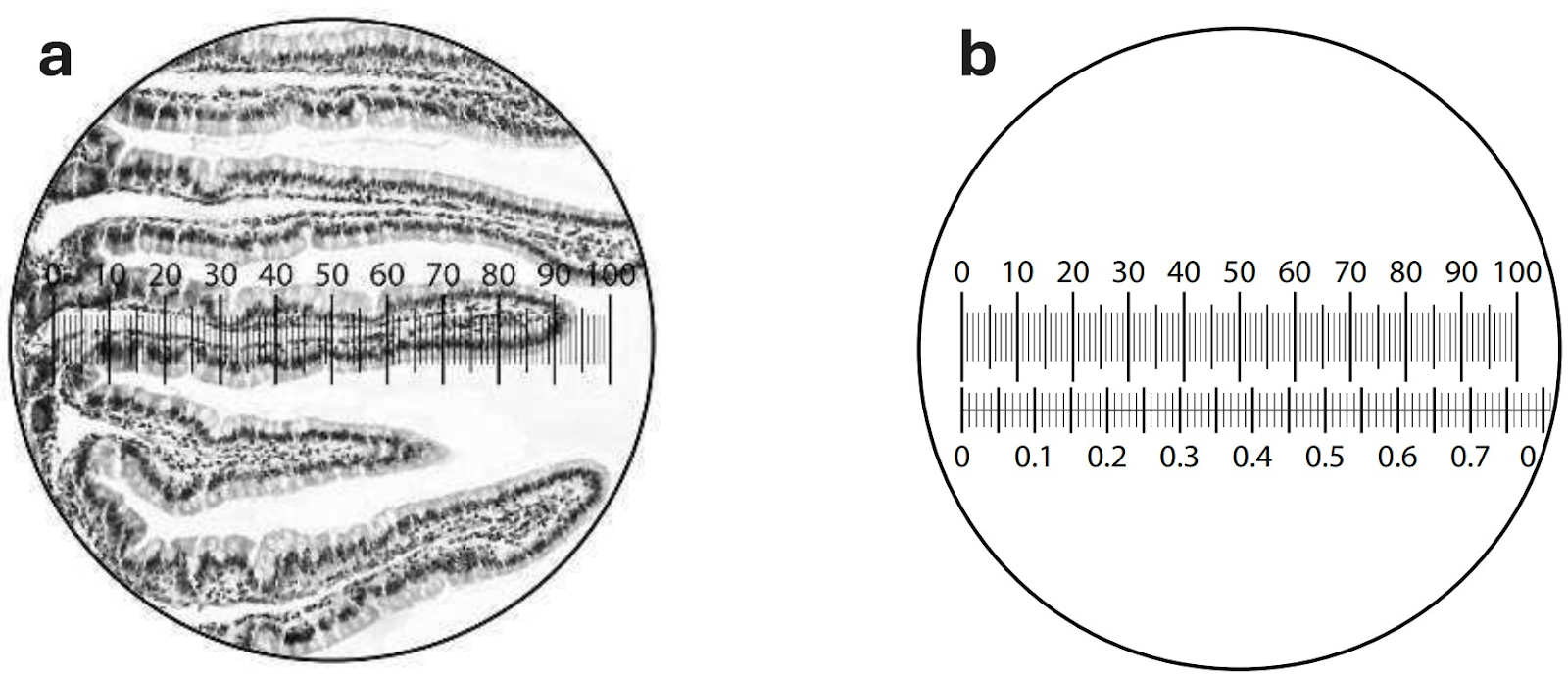
How long is the villus that can be seen beneath the eyepiece graticule?
A. `6.81 ×10^2 µm`
B. `6.81 ×10^-2 µm`
C. `681 ×10^2 mm`
D. `681 ×10^-2 mm`
Medium
Mark as Complete
Mark Scheme
Question 4
What magnification occurs in a light microscope with `a ×6` eyepiece lens and `a ×10` objective lens?
Easy
Mark as Complete
Mark Scheme
Question 5
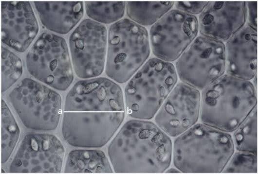
The figure below shows a micrograph of plant cells. The actual length of the cell between points a and b on the line is `12 µm`. The length of the line between a and b on the diagram is `3 mm`. Calculate the magnification of this image.
Easy
Mark as Complete
Mark Scheme
Question 6
The table below shows the comparison of light microscopes and electron microscopes. Some boxes have been filled in for you. Copy and complete the table.
| Feature | Light microscope | Electron microscope |
| source of radiation | ||
| wavelength of radiation | about 0.005 nm | |
| maximum resolution | 0.5 nm in practice | |
| lenses | glass | |
| specimen | non-living or dead | |
| stains | coloured dyes | |
| image | coloured |
Medium
Mark as Complete
Mark Scheme
Question 7
Copy and complete this table. In the ‘type of microscope’ column, choose from optical microscope, transmission electron microscope or scanning electron microscope.
| Micrograph | Type of microscope used to produce the micrograph | Reason for your decision |
|
Figure a. Micrograph of plant cells with starch grains. | ||
|
| ||
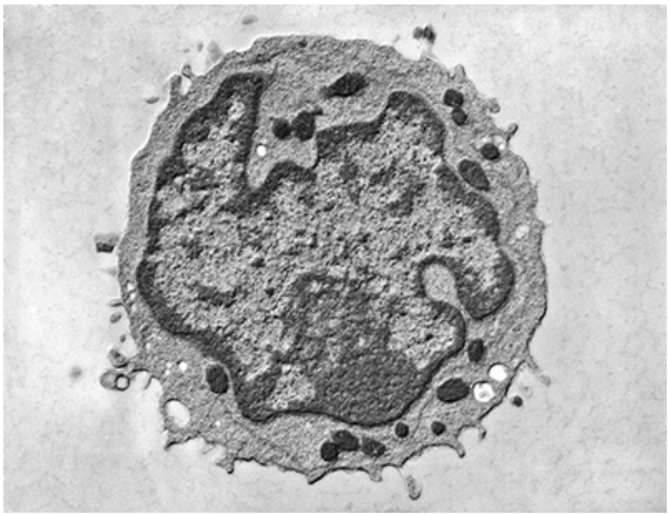 Figure c. Micrograph of lymphocyte. Figure c. Micrograph of lymphocyte. |
Medium
Mark as Complete
Mark Scheme
Question 8
Explain why the nucleus in an animal cell may be viewed by light microscopy in an appropriately stained cell but the ribosomes cannot.
Easy
Mark as Complete
Mark Scheme
Question 9
Copy and complete this table, to compare what can be seen in typical animal cells and plant cells using optical microscopes and electron microscopes. Put a tick or a cross in each box.
| Organelle | Visible in plant cells | Visible in plant cells | ||
| Visible using optical microscope | Visible using electron microscope | Visible using optical microscope | Visible using electron microscope | |
| nucleus | ||||
| mitochondrion | ||||
| membranes within mitochondrion | ||||
| Golgi body | ||||
| ribosomes | ||||
| endoplasmic reticulum | ||||
| chloroplast | ||||
| internal structure of chloroplast | ||||
| centriole | ||||
Medium
Mark as Complete
Mark Scheme
Question 10
The figure below shows three fields of view seen using a high-power (×40) objective lens: a an eyepiece graticule scale; b human cheek epithelial cells and the eyepiece graticule scale; c the eyepiece graticule scale and the stage micrometer scale. Calibrate the eyepiece graticule to then calculate the actual diameter of the cell shown on the eyepiece graticule.
Hard
Mark as Complete
Mark Scheme
Question 1
Identify the correct statement about a eukaryotic cell.
A. Eukaryotic cells have no membrane-bound nuclei.
B. Prokaryotic cells evolved from eukaryotic cells.
C. Eukaryotic cells include animals, plants, fungi and some other organisms.
D. Eukaryotic cells contain fewer different organelles than prokaryotic cells.
Answer: C
A. Incorrect: The eukaryotic cell has a nucleus, which is a membrane-bound organelle. The nucleus is surrounded by a nuclear envelope, which is a double membrane. The prokaryotic cell, in contrast, lacks a membrane-bound nucleus.
B. Incorrect: Prokaryotic cells appeared on Earth much earlier than eukaryotic cells. Eukaryotes evolved from prokaryotes. The theory of endosymbiosis suggests a possible mechanism for this evolution, proposing that organelles like mitochondria and chloroplasts originated as free-living prokaryotes that were taken up by a host cell and came to survive as organelles within it.
C. Correct: Animals, plants, and fungi are organisms made up of eukaryotic cells. Protoctists are also eukaryotes, including organisms like algae and protozoa.
D. Incorrect: Eukaryotic cells are generally more complicated and having more organelles than prokaryotic cells. Prokaryotic cells are simpler and much smaller, lacking the membrane-bound organelles found in the cytoplasm of eukaryotes. The cytoplasm of eukaryotic cells contains numerous organelles, many of which are membrane-bound structures like the nucleus, mitochondria, chloroplasts, endoplasmic reticulum, Golgi apparatus, and lysosomes.
Question 2
Calculate how many millimetres there are in `2.3` nanometres.
A. `2300``mm`
B. `2 300 000` `mm`
C. `0.0023` `mm`
D. `0.000 0023` `mm`
Answer: D
| Unit | Standard form for metres |
| nanometres `(nm)` | `X10-9 m` |
| micrometres `(µm)` | `X 10-6 m` |
| millimetres `(mm)` | `X 10-3 m` |
There are `1000` `nm` in `1` µm, or`1` `nm` =`0.001 µm`
There are`1000 µm` in`1` `mm`, or `1 µm` = `0.001 mm`
So: `1 nm = 0.000001 mm`
`2.3 nm = 2.3×0.000001 mm`
`= 0.0000023 mm`
Question 3
Figure a shows a light micrograph of some villi in the small intestine, seen using an eyepiece graticule. Figure b shows the same eyepiece graticule, using the same objective lens, but this time with a stage micrometer on the microscope stage.
How long is the villus that can be seen beneath the eyepiece graticule?
A. `6.81 ×10^2 µm`
B. `6.81 ×10^-2 µm`
C. `681 ×10^2 mm`
D. `681 ×10^-2 mm`
Answer: A
In Figure a, the `5` mark on the eyepiece graticule scale is at the bottom of the villus, and the `94` mark at the tip. The villus therefore measures `94 − 5 = 89` eyepiece units.
In Figure b, `100` eyepiece graticule units measure `76.5` micrometer units. As each small micrometer mark represents `0.01 mm`, `76.5`micrometer units `= 0.765 mm`.
So `89` eyepiece units represent:
`89/100×0.765=0.681 mm`
Converting mm to µm:
`0.681 mm ×1000=681 µm= 6.8110^2 µm`
Question 4
What magnification occurs in a light microscope with `a ×6` eyepiece lens and `a ×10` objective lens?
The total magnification in a light microscope is calculated by multiplying the magnification of the eyepiece lens by the magnification of the objective lens.
Total magnification = Eyepiece magnification × Objective lens magnification
`= 6 × 10`
`= ×60`
The magnification that occurs in a light microscope with `a ×6` eyepiece lens and `a ×10` objective lens is `×60`.
The multiplication sign `(×)` in front of the number `60` means 'times'. We say that the magnification is 'times `60'`.
Question 5
The figure below shows a micrograph of plant cells. The actual length of the cell between points a and b on the line is `12 µm`. The length of the line between a and b on the diagram is `3 mm`. Calculate the magnification of this image.
The magnification of the image:
Magnification, `M=frac{"Image size",I}{"Actual size", A}`
(Note that the units for the size of the image and the actual size of the specimen are the same)
Substitute the values into the magnification formula:
`"Magnification"=frac{"Image size"}{"Actual size"}=frac{3000 µm}{12 µm}=×250`
The magnification of this image is `×250`.
Question 6
The table below shows the comparison of light microscopes and electron microscopes. Some boxes have been filled in for you. Copy and complete the table.
| Feature | Light microscope | Electron microscope |
| source of radiation | ||
| wavelength of radiation | about 0.005 nm | |
| maximum resolution | 0.5 nm in practice | |
| lenses | glass | |
| specimen | non-living or dead | |
| stains | coloured dyes | |
| image | coloured |
| Feature | Light microscope | Electron microscope |
| source of radiation | light | electrons |
| wavelength of radiation | 400-700 nm | about 0.005 nm |
| maximum resolution | 200 nm | 0.5 nm in practice |
| lenses | glass | electromagnets |
| specimen | living, non-living or dead | non-living or dead |
| stains | coloured dyes | heavy metals |
| image | coloured | black and white |
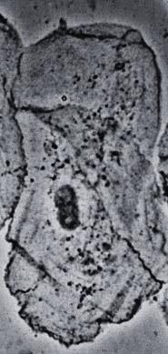
Question 7
Copy and complete this table. In the ‘type of microscope’ column, choose from optical microscope, transmission electron microscope or scanning electron microscope.
| Micrograph | Type of microscope used to produce the micrograph | Reason for your decision |
|
Figure a. Micrograph of plant cells with starch grains. | ||
|
| ||
| Figure c. Micrograph of lymphocyte. |
Scanning electron microscopes (SEMs) scan a beam of electrons across the specimen, producing images that show the surface and can be 3D. Transmission electron microscopes (TEMs) transmit a beam of electrons through a thin specimen, giving high resolution images that show the internal structure of organelles and are in 2D. Optical (light) microscopes use light and glass lenses and have lower resolution and magnification limits compared to electron microscopes. While light microscopes typically show less detail than electron microscopes, a very good one might show some internal nuclear detail like chromatin distribution
| Micrograph | Type of microscope used to produce the micrograph | Reason for your decision |
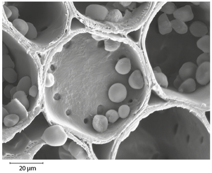 Figure a. Micrograph of plant cells with starch grains. Figure a. Micrograph of plant cells with starch grains. | scanning electron microscope (SEM) | in 3D and shows detail more clearly than would be visible using an optical microscope |
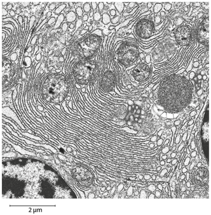 Figure b. Micrograph of a cell from the pancreas. Figure b. Micrograph of a cell from the pancreas. | transmission electron microscope (TEM) | in 2D and has very high resolution, e.g. membranes of endoplasmic reticulum can be clearly seen |
| Figure c. Micrograph of lymphocyte. | could be very good optical microscope or transmission electron microscope | in 2D; this amount of detail, including the distribution of dark and light-staining chromatin in the nucleus, could be visible using a very good optical microscope |
Question 8
Explain why the nucleus in an animal cell may be viewed by light microscopy in an appropriately stained cell but the ribosomes cannot.
The ability to distinguish between two separate points using a light microscope is limited by its resolution (maximum `200 nm`). Staining is necessary to provide contrast between the cytosol and the nucleus in an otherwise more or less transparent cell. The nucleus is a relatively large organelle in eukaryotic cells (`10-20 µm`), which is visible when using a light microscope. Ribosomes, however, are too small to be resolved by a light microscope, regardless of staining.
Question 9
Copy and complete this table, to compare what can be seen in typical animal cells and plant cells using optical microscopes and electron microscopes. Put a tick or a cross in each box.
| Organelle | Visible in plant cells | Visible in plant cells | ||
| Visible using optical microscope | Visible using electron microscope | Visible using optical microscope | Visible using electron microscope | |
| nucleus | ||||
| mitochondrion | ||||
| membranes within mitochondrion | ||||
| Golgi body | ||||
| ribosomes | ||||
| endoplasmic reticulum | ||||
| chloroplast | ||||
| internal structure of chloroplast | ||||
| centriole | ||||
| Organelle | Visible in plant cells | Visible in animal cells | ||
| Visible using optical microscope | Visible using electron microscope | Visible using optical microscope | Visible using electron microscope | |
| nucleus | ✓ | ✓ | ✓ | ✓ |
| mitochondrion | ✓ | ✓ | ✓ | ✓ |
| membranes within mitochondrion | ✗ | ✓ | ✗ | ✓ |
| Golgi body | ✓ | ✓ | ✓ | ✓ |
| ribosomes | ✗ | ✓ | ✗ | ✓ |
| endoplasmic reticulum | ✗ | ✓ | ✗ | ✓ |
| chloroplast | ✓ | ✓ | ✗ | ✗ |
| internal structure of chloroplast | ✗ | ✓ | ✗ | ✗ |
| centriole | ✗ | ✗ | ✓ | ✓ |
Question 10
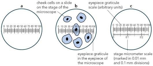
The figure below shows three fields of view seen using a high-power (×40) objective lens: a an eyepiece graticule scale; b human cheek epithelial cells and the eyepiece graticule scale; c the eyepiece graticule scale and the stage micrometer scale. Calibrate the eyepiece graticule to then calculate the actual diameter of the cell shown on the eyepiece graticule.
Cells and organelles can be measured with a microscope by means of an eyepiece graticule. This is a transparent scale. It usually has `100` divisions.
To calibrate the eyepiece graticule scale, a miniature transparent ruler called a stage micrometer scale is placed on the microscope stage and is brought into focus. It commonly has subdivisions of `0.1` and `0.01 mm`.
In the eyepiece graticule shown, `100` eyepiece units measure `v`on the stage micrometer.
The value of one eyepiece unit:
`frac(0.25 mm)(100)=0.0025 mm`
Converting mm to µm:
`0.0025 mm ×1000=0.0025 µm`
The cell spans from `40` to `60` on the eyepiece scale, so the diameter of the cell measures `20` eyepiece units.
The actual diameter:
`20 ×2.5 μm=50 μm`
This diameter is greater than that of many human cells because the cell is a flattened epithelial cell.
Question 1
Identify the correct statement about a eukaryotic cell.
A. Eukaryotic cells have no membrane-bound nuclei.
B. Prokaryotic cells evolved from eukaryotic cells.
C. Eukaryotic cells include animals, plants, fungi and some other organisms.
D. Eukaryotic cells contain fewer different organelles than prokaryotic cells.
Question 2
Calculate how many millimetres there are in `2.3` nanometres.
A. `2300``mm`
B. `2 300 000` `mm`
C. `0.0023` `mm`
D. `0.000 0023` `mm`
Question 3
Figure a shows a light micrograph of some villi in the small intestine, seen using an eyepiece graticule. Figure b shows the same eyepiece graticule, using the same objective lens, but this time with a stage micrometer on the microscope stage.
How long is the villus that can be seen beneath the eyepiece graticule?
A. `6.81 ×10^2 µm`
B. `6.81 ×10^-2 µm`
C. `681 ×10^2 mm`
D. `681 ×10^-2 mm`
Question 4
What magnification occurs in a light microscope with `a ×6` eyepiece lens and `a ×10` objective lens?
Question 5
The figure below shows a micrograph of plant cells. The actual length of the cell between points a and b on the line is `12 µm`. The length of the line between a and b on the diagram is `3 mm`. Calculate the magnification of this image.
Question 6
The table below shows the comparison of light microscopes and electron microscopes. Some boxes have been filled in for you. Copy and complete the table.
| Feature | Light microscope | Electron microscope |
| source of radiation | ||
| wavelength of radiation | about 0.005 nm | |
| maximum resolution | 0.5 nm in practice | |
| lenses | glass | |
| specimen | non-living or dead | |
| stains | coloured dyes | |
| image | coloured |
Question 7
Copy and complete this table. In the ‘type of microscope’ column, choose from optical microscope, transmission electron microscope or scanning electron microscope.
| Micrograph | Type of microscope used to produce the micrograph | Reason for your decision |
|
Figure a. Micrograph of plant cells with starch grains. | ||
|
| ||
| Figure c. Micrograph of lymphocyte. |
Question 8
Explain why the nucleus in an animal cell may be viewed by light microscopy in an appropriately stained cell but the ribosomes cannot.
Question 9
Copy and complete this table, to compare what can be seen in typical animal cells and plant cells using optical microscopes and electron microscopes. Put a tick or a cross in each box.
| Organelle | Visible in plant cells | Visible in plant cells | ||
| Visible using optical microscope | Visible using electron microscope | Visible using optical microscope | Visible using electron microscope | |
| nucleus | ||||
| mitochondrion | ||||
| membranes within mitochondrion | ||||
| Golgi body | ||||
| ribosomes | ||||
| endoplasmic reticulum | ||||
| chloroplast | ||||
| internal structure of chloroplast | ||||
| centriole | ||||
Question 10
The figure below shows three fields of view seen using a high-power (×40) objective lens: a an eyepiece graticule scale; b human cheek epithelial cells and the eyepiece graticule scale; c the eyepiece graticule scale and the stage micrometer scale. Calibrate the eyepiece graticule to then calculate the actual diameter of the cell shown on the eyepiece graticule.

